

This advanced procedure helps stop the progression of keratoconus.
Advanced corneal collagen cross-linking uses ultraviolet (UV) light with vitamin B2 (riboflavin) eye drops to strengthen corneal tissue. The treatment can slow or halt the progression of keratoconus. Led by Newsom Eye founder and Medical Director, T. Hunter Newsom, MD, Newsom Eye is proud to offer this treatment option to patients with keratoconus, providing them with NEWSOM EYES!
What is Keratoconus?
Keratoconus is an abnormality of the eye in which the normally round, dome-shaped cornea weakens and becomes progressively thinner and irregular in shape. The resulting cone-shaped cornea can cause high levels of astigmatism (irregular curvature) and nearsightedness. Keratoconus has been estimated to occur in one out of every 1,000 persons in the general population and is generally first diagnosed in young people at puberty or in their late teens. Prior to collagen cross-linking, there was no way to stop the progression of this disease.
What Is Collagen Cross-Linking?
Corneal collagen cross-linking (CXL), a procedure first developed in Dresden, Germany, in the 1990s, is a technique used to treat individuals with progressive keratoconus. This is the only procedure that can strengthen the cornea to slow or stop the progression of keratoconus. This minimally invasive treatment utilizes Vitamin B2 (riboflavin) and ultraviolet (UVA) light to increase the cornea’s collagen connections (cross-links), which helps the cornea retain its firmness and shape. Collagen cross-linking is not a cure for keratoconus.
How Does Collagen Cross-Linking Work?
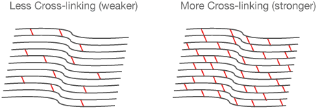 Collagen is the structural protein that provides strength and firmness to the corneal tissue. CXL works by forming new cross-links between collagen fibers, which are the natural “anchors” within the cornea. These anchors are responsible for preventing the weakened cornea from bulging out and becoming steep and irregular. The addition of these “cross beams” acts like adding rungs on a ladder to stiffen and strengthen the cornea, allowing it to focus light more efficiently and accurately.
How Is Collagen Cross-Linking Performed?
The collagen cross-linking treatment is an outpatient procedure performed in our office. First, you will be given anesthetic (numbing) eye drops to prevent any pain or discomfort. Then, Vitamin B2 (riboflavin) eye drops are put in the eye, one drop every two minutes for about 45 minutes. To confirm that the treatment will be effective, the doctor will check your eye to make sure that the Vitamin B2 (riboflavin) drops have saturated the cornea. Next, you will relax in a reclining chair and look up at a soft blue ultraviolet light (UVA) during treatment. Also, to help prevent blinking, an eyelid retainer will be placed between the upper and lower eyelids while the ultraviolet light source is then shined into your eye. The light “activates” the Vitamin B2 (riboflavin), which causes the cross-linking of the collagen fibers. The UVA treatment will last approximately five to 10 minutes, and it generally is not difficult to look at the light because your eyes are numb. A clear, soft, and powerless contact lens may be placed on your eye immediately after the procedure, where it will remain in place for several days to aid healing and comfort.
How Effective Is Collagen Cross-Linking and Does it Improve Vision?
By strengthening collagen links, CXL is the first keratoconus treatment that goes to the root of the disease. Many research studies have shown that collagen cross-linking prevents further vision loss in more than 95% of patients. Research has also shown that more than 50% of patients saw some overall improvement in vision. As with many conditions, the earlier the diagnosis, the better the results. If collagen cross-linking is performed early, it may be possible to prevent the signs and symptoms of keratoconus.