Retina Care

Retina Care in Tampa, Sebring, Brooksville & Gainesville

You deserve NEWSOM EYES!

The retina is the film of the eye. New floaters, flashes of light or a shadow across your vision are never something to wait on. Newsom Eye examines and images the retina at all five Florida locations.


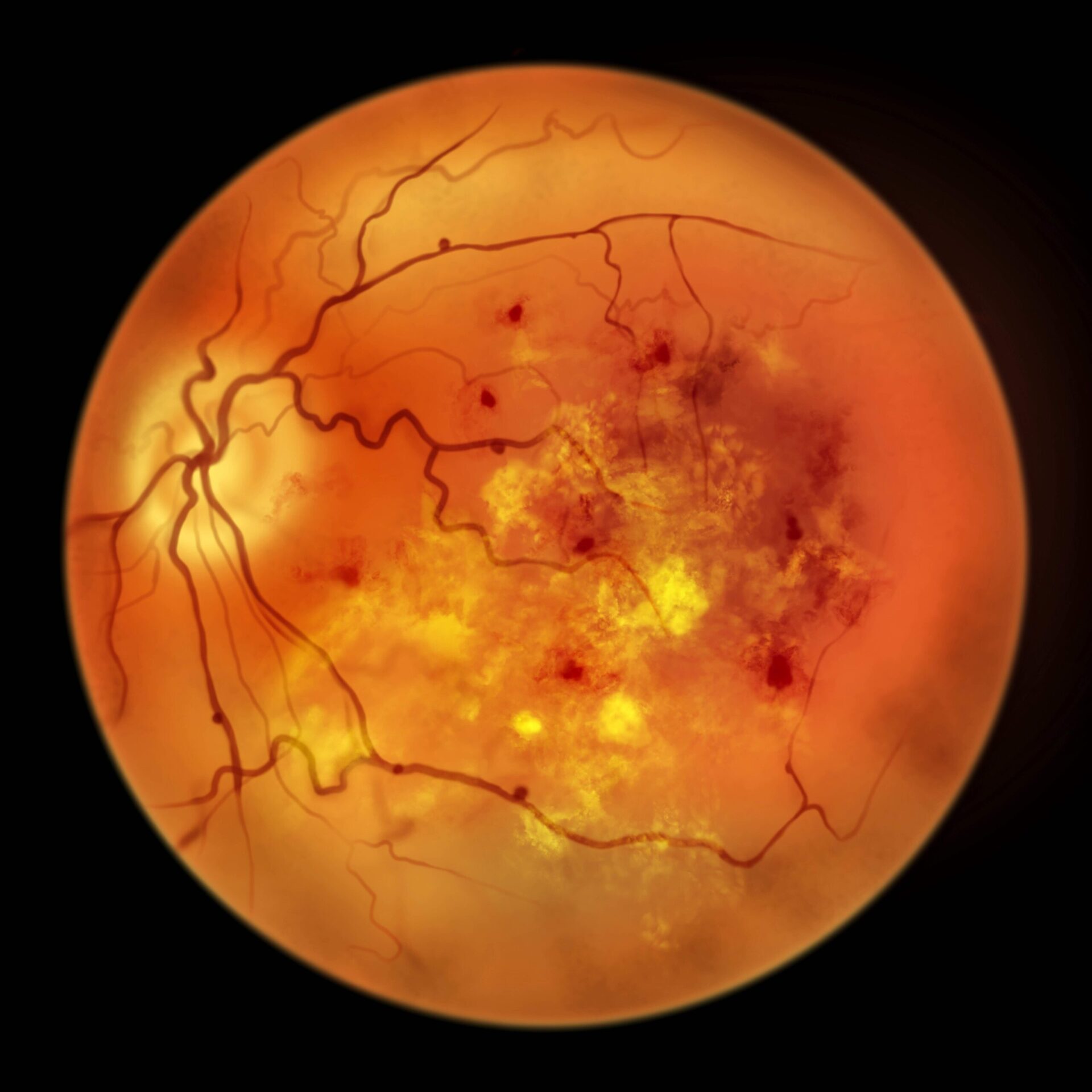
Retinal imaging of the back of the eye at Newsom Eye
25+ Years
Serving Florida patients
100,000+
Surgeries performed
5 Locations
Sebring to Gainesville
3 Surgery Centers
AAAHC accredited
Anatomy of the retina and macula at the back of the eye
The Basics

How does the retina function?

The retina is the thin layer of tissue that lines the back of the eye. It functions like the film in a camera — capturing light and processing it into the information your brain assembles into the images you see.

The macula is the most specialized part of the retina and home to the sensitive photoreceptors that let you focus on fine detail. There are two types of photoreceptor cells, rods and cones. Without them working properly, even simple tasks — reading, driving, watching television — become difficult.

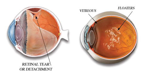
Retinal health is one of the most important factors affecting eyesight today. Patients often describe objects floating through their field of vision, or sudden lightning-like flashes of light. Most of these are common and minor, but left unchecked they can lead to retinal tears and detachment.

  • Rods and cones. Photoreceptors that turn light into signals your brain reads.
  • The macula. The small central area responsible for sharp, detailed vision.
  • No spare parts. Retinal tissue that dies does not grow back — timing is everything.
Simulation of flashes and floaters in the field of vision
Do Not Wait

Retinal warning signs that need same-day attention

Most floaters are harmless and settle over time. What matters is change. A sudden shower of new floaters, repeated flashes of light, or a dark curtain moving across your vision are the classic signs of a retinal tear or detachment.

Caught as a tear, the retina can often be sealed in the office with a laser. Once it has detached, repair means surgery — and the vision that comes back depends heavily on how quickly it was treated.

If you notice any of these symptoms, call (813) 908-2020 and tell us what you are seeing. Do not book a routine appointment and wait.

  • A sudden shower of new floaters where before there were few.
  • Repeated flashes of light, often at the edge of vision.
  • A curtain or shadow moving across part of your vision.
  • Any sudden loss of vision, even if it seems to improve.
What to Expect

How Newsom Eye examines the retina

A retinal evaluation is painless, takes about an hour with dilation, and produces images we can compare year over year.

1

Dilation

Drops widen the pupil so the entire retina, right out to its edges, can be examined rather than just the central view.

2

Wide-angle retinal imaging

A high-resolution photograph of the retina that documents its condition today and becomes the baseline for every future visit.

3

OCT scanning

A cross-section of the retinal layers that shows swelling, fluid and macular changes long before they affect vision.

4

Diagnosis and staging

Your doctor reviews the images with you and explains what is present, what is stable and what needs to be watched.

5

Treatment or referral

In-office laser for tears, monitoring and nutritional guidance for dry AMD, and prompt coordination with a retina surgeon when a detachment or injection therapy is needed.

6

Scheduled follow-up

Retinal disease is tracked with repeat imaging. Comparing scans is how progression gets caught while treatment still works.

#1AMD is the top cause of vision loss over 60
Age 50+When annual dilated exams should start
1 YearMaximum interval between exams with diabetes
Same DayHow fast a curtain or shadow should be seen
Patient Stories

Real Newsom Eye patients

Meet Alejandra: A Vision of SuccessMeet Alejandra: A Vision of Success
Amazing Cataract OutcomesAmazing Cataract Outcomes
Amazing Glaucoma OutcomesAmazing Glaucoma Outcomes
Retina FAQs

Frequently asked questions about the retina

What does the retina do?

The retina is the thin layer of tissue lining the back of the eye. It works like the film in a camera — capturing light and converting it into signals your brain assembles into images. The macula, at its center, holds the photoreceptors responsible for sharp detail vision used for reading and driving.

What are flashes and floaters?

Floaters are tiny clumps of cells or gel inside the vitreous, the clear jelly that fills the eye. They appear as specks, threads or clouds drifting through your vision. Flashes are brief arcs or streaks of light caused by the vitreous tugging on the retina. Most are harmless, but a sudden increase in either can signal a retinal tear.

When are flashes and floaters an emergency?

Call us the same day if you notice a sudden shower of new floaters, repeated flashes of light, a dark curtain or shadow moving across your vision, or a sudden drop in vision. Those are the warning signs of a retinal tear or detachment, and treatment is far more effective before the retina fully detaches.

What is a retinal detachment?

A detachment occurs when the retina separates from the wall of the eye and loses its blood supply. Without prompt repair, the affected retina stops working permanently. It is one of the few true emergencies in eye care.

What is macular degeneration?

Age-related macular degeneration (AMD) is the leading cause of vision loss among Americans over 60. It damages the macula and takes central vision while leaving peripheral vision intact. Risk factors include age, family history and smoking.

Can macular degeneration be treated?

Dry AMD is managed with monitoring, specific vitamin formulations and lifestyle changes such as quitting smoking. Wet AMD is treated with anti-VEGF injections that can stabilize and sometimes improve vision. Both depend on catching the disease early, which is why annual dilated exams after age 50 matter.

How does diabetes affect the eyes?

Diabetes damages the small blood vessels of the retina, causing them to leak or close off. Diabetic retinopathy often has no symptoms in its early, most treatable stages. Everyone with diabetes should have a dilated retinal exam at least once a year.

How is the retina examined?

Through a dilated pupil, using wide-angle retinal imaging and OCT scanning that photographs and cross-sections the retinal layers. These images also create a baseline, so future changes can be compared rather than guessed at.

Where does Newsom Eye evaluate retinal problems?

Retinal examination and imaging are available at all five Newsom Eye locations — Sebring, Carrollwood (Tampa), South Tampa, Brooksville and Gainesville. If you have symptoms of a tear or detachment, call us immediately rather than waiting for a routine appointment.

Protect your retina before symptoms start

Macular degeneration and diabetic eye disease are silent in their earliest and most treatable stages. Schedule a dilated retinal exam at any of our five Florida locations.